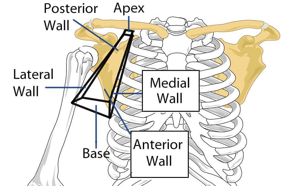
The axilla or armpit is a pyramidal space situated between the upper part of the arm and the chest wall. It
resembles a four-sided pyramid, and has
(i) an apex
(ii) a base
(iii) 4 walls—anterior, posterior, medial and
lateral
Its apex is bound superiorly by the root of the neck. The apex is also referred to as the axillary inlet. The borders of the apex are made by the lateral surface of the first rib, the posterior surface of the clavicle, and the superior margin of the scapula.
- The medial border is created by the serratus anterior, as well as the thoracic wall which includes intercostal muscles and costals (ribs) of that region.
- The lateral border is made by the intertubercular sulcus (groove) of the humerus.
- The floor, or base, of the axilla is the axillary fascia and its skin on the surface of the axilla, aka the ‘armpit’. The axilla is filled with adipose (fat) and allows passage for: vessels, nerve plexus, lymphatics, and muscles. The muscles include the coracobrachialis, pectoralis minor, and the biceps brachii.
- The posterior border of pectoralis major and minor defines the anterior border (fold).
- The posterior border (fold) is the subscapularis superiorly, and the latissimus dorsi and teres major inferiorly. The intercostobrachial nerves supply sensation to the skin of the axilla.
Contents
The contents of the axilla region include muscles, nerves, vessels, and lymphatics:
- Axillary artery (and branches) – the main artery supplying the upper limb. It is commonly referred as having three parts; one medial to the pectoralis minor, one posterior to pectoralis minor, and one lateral to pectoralis minor. The medial and posterior parts travel in the axilla.
- Axillary vein (and tributaries) – the main vein draining the upper limb, its two largest tributaries are the cephalic and basilic veins.
- Brachial plexus (and branches) – a collection of spinal nerves that form the peripheral nerves of the upper limb.
- Axillary lymph nodes – they filter lymphatic fluid that has drained from the upper limb and pectoral region. Axillary lymph node enlargement is a non-specific indicator of breast cancer.
- Biceps brachii (short head) and coracobrachialis – these muscle tendons move through the axilla, where they attach to the coracoid process of the scapula.
Clinical Anatomy
Thoracic Outlet Syndrome
The apex of the axilla region is an opening between the clavicle, first rib and the scapula. In this apex, the vessels and nerves may become compressed between the bones – this is called thoracic outlet syndrome.
Common causes of thoracic outlet syndrome include:
- Trauma – e.g. fractured clavicle.
- Repetitive movements – seen commonly in occupations that require lifting of the arms.
- Cervical rib – an extra rib which arises from the seventh cervical vertebra.
It often presents with pain in the affected limb (the distribution of pain is dependent on which nerve is compressed), tingling, muscle weakness and discolouration.
Lymph Node Biopsy
Approximately 75% of lymph from the breast drains into the axilla lymph nodes, so can be biopsied if breast cancer is suspected.
If breast cancer is confirmed, the axillary nodes may need to be removed to prevent the cancer spreading. This is known as axillary clearance. During this procedure, the long thoracic nerve may become damaged, resulting in winged scapula.